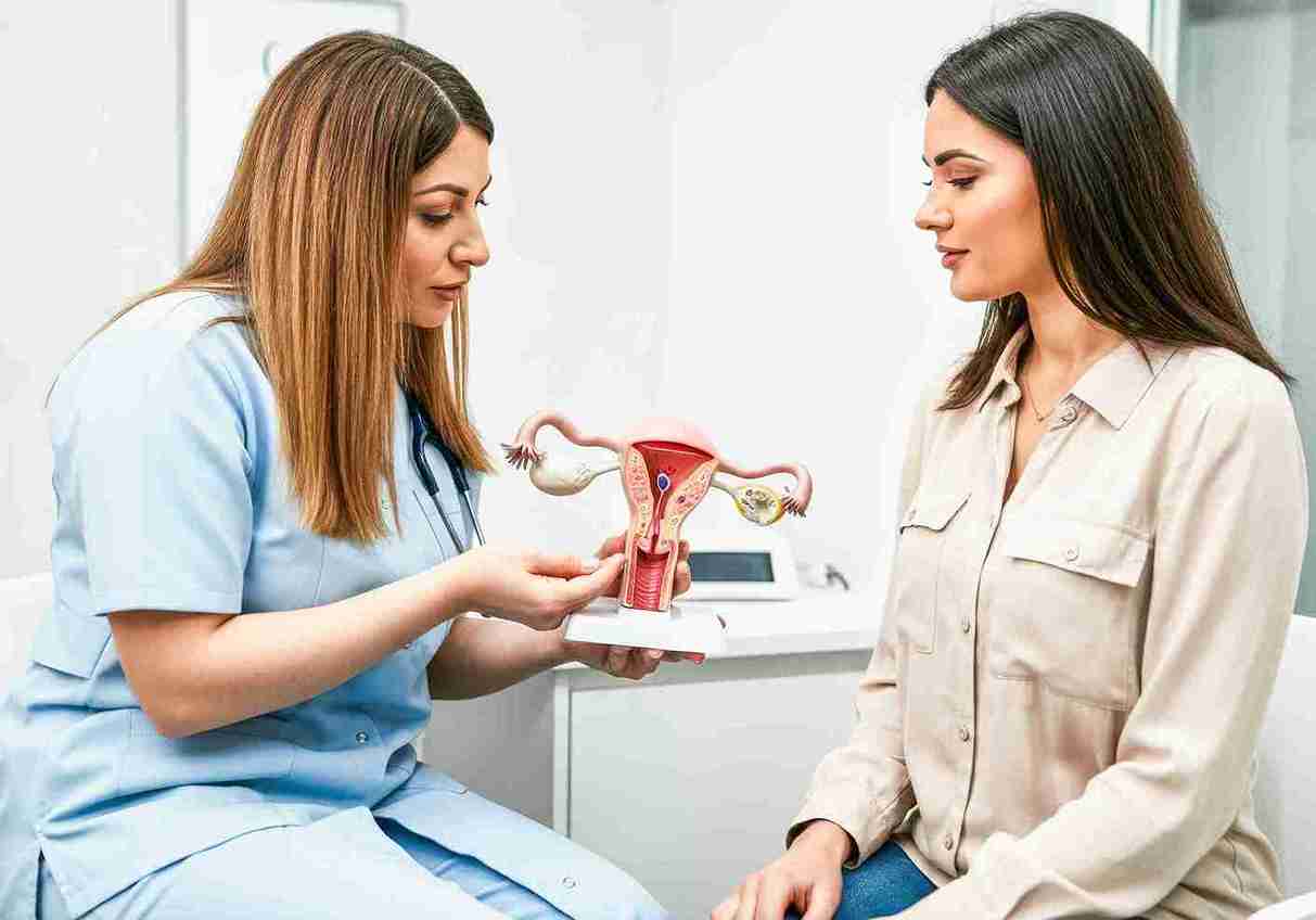
A hysteroscopy is a short-daycare procedure in which a thin camera is passed through the cervix to look inside the uterus, and to treat problems in the same sitting. In fertility care, it earns its place when it finds and fixes a real abnormality - a polyp, fibroid, scar tissue, or septum. Routinely scoping a normal-looking cavity, by contrast, does not improve IVF success.
What Hysteroscopy Is - Diagnostic vs Operative
Hysteroscopy (a camera examination of the inside of the uterus) uses a narrow telescope-like instrument, the hysteroscope, passed through the cervix while a little fluid gently opens up the cavity so it can be seen clearly. There are two forms, and the difference matters for what to expect:
Type
What it does
Setting
Diagnostic
Looks inside the uterus to confirm or rule out a problem
Usually outpatient or short day-care; quick recovery
Operative
Treats what is found - removes a polyp, fibroid, or scar tissue, or corrects a septum
Day-care, often under sedation or short general anaesthesia
Often, both happen together: the specialist looks and, if a treatable problem is found, corrects it in the same procedure - a “see-and-treat” approach that saves a second visit.
How It Is Done in India - Anaesthesia, Day-Care and Time Off
In many Western centres, a diagnostic hysteroscopy is done in the clinic under local anaesthesia or none at all. In India, practice leans towards comfort: it is commonly performed as a day-care procedure under sedation or a short general anaesthetic, particularly when treatment is likely in the same sitting. You are usually admitted in the morning and discharged the same day. For a straightforward diagnostic hysteroscopy, most women take just the day off and return to work the next day; an operative procedure that corrects a significant abnormality may require a couple of days. It is worth confirming the anaesthesia plan and expected time off with your centre in advance, so you can arrange leave and someone to accompany you home.
When is Hysteroscopy Recommended - and Does It Help Before IVF?
When it is recommended
Hysteroscopy is not a routine test for everyone trying to conceive. It is recommended when there is a specific reason to look inside the uterus - an abnormal ultrasound or HSG test, unexplained abnormal bleeding, suspected polyps or fibroids, recurrent miscarriage, or repeated IVF failure, where a uterine cause needs ruling in or out.
Does it improve IVF success? The honest answer
This is where careful, evidence-based framing matters. Two large randomised trials settled a long-running debate: in women whose uterine cavity looks normal on ultrasound, performing a hysteroscopy before IVF does not improve the live-birth rate, whether before a first cycle or after failed cycles. What does help is hysteroscopy, which finds and corrects a genuine problem: removing a polyp, a submucosal fibroid, or scar tissue improves the chance of a pregnancy. Since an intrauterine abnormality is present in roughly a quarter of women being assessed for infertility, the value lies in detection and treatment - not in scoping a healthy cavity for reassurance.
After two or more failed IVF cycles
This is a common and reasonable trigger. After repeated implantation failure (two or more IVF transfers that did not result in pregnancy), a hysteroscopy can find subtle problems that scans miss - small polyps, mild scar tissue, or chronic inflammation of the lining (chronic endometritis). If something is found and treated, that is worthwhile. If the cavity is genuinely normal, the honest expectation is that the hysteroscopy itself will not lift your odds, and attention should turn to other factors, such as embryo quality or the transfer protocol.
What Hysteroscopy Reveals That a Scan Can Miss
A standard ultrasound is a good first look, but it cannot see the cavity in the detail a camera can. Hysteroscopy can find and, in many cases, treat:
Endometrial polyps: soft overgrowths of the lining that can interfere with implantation - often missed or under-called on a routine scan.
Submucosal fibroids: fibroids that bulge into the cavity itself, distorting the space where an embryo would implant.
Intrauterine adhesions (Asherman’s syndrome): bands of scar tissue, often after a previous uterine procedure or infection, that can stick the walls together.
Uterine septum: a band of tissue dividing the cavity, present from birth; whether to correct it is individualised, as the evidence is debated.
Chronic endometritis: low-grade, silent inflammation of the lining that a scan cannot show.
Retained tissue: remnants from a previous pregnancy or procedure that need clearing under direct vision.
How the Procedure Works and What Recovery Looks Like
Step by step
You are admitted as a day-care patient; anaesthesia (local, sedation, or a short general) is arranged for comfort.
The hysteroscope is passed gently through the vagina and cervix - no cuts are made on the abdomen.
A small amount of fluid opens the cavity, allowing the lining and tube openings to be seen clearly on a screen.
If a treatable problem is found, fine instruments passed through the scope remove or correct it in the same sitting.
The whole procedure usually takes 15-30 minutes, and you are discharged the same day.
Recovery and getting back to work
Expect mild, period-like cramping and light spotting for a day or two. After a diagnostic hysteroscopy, most women return to normal activity the next day. After an operative procedure that corrected something significant - a large fibroid, extensive scar tissue, or a septum - recovery can take a little longer, and specialists often advise waiting roughly one to three months before trying to conceive or proceeding to a frozen embryo transfer (FET), to let the lining heal. Your own timeline depends on what was done, so follow your specialist’s specific advice.
Is Hysteroscopy Right for You?
Whether a hysteroscopy is likely to help depends on your situation:
Your situation
Is hysteroscopy likely indicated?
Why
Abnormal scan or HSG suggesting a polyp, fibroid, or adhesion
Yes
To confirm and treat a real, identified problem
Recurrent miscarriage
Often
To check for septum, adhesions, or chronic inflammation
Repeated IVF failure with a normal cavity
Selective
Useful to find missed pathology; no benefit if the cavity is truly normal
Routine, before a first IVF, normal cavity
Usually no
Trial evidence shows no live-birth benefit
Heavy or irregular bleeding is being investigated
Yes
Direct view and targeted sampling
If a scan, an HSG test, or repeated IVF treatment setbacks have raised a question about your uterus, you can book a fertility consultation at a Cloudnine Fertility centre to find out whether a hysteroscopy is the right next step for you.
Treating a polyp / fibroid / adhesion found at hysteroscopy
Improves the chance of pregnancy
Reviews of operative hysteroscopy
Moderate
How often is a cavity problem present in infertility
About 1 in 4 women assessed
Cohort data
Moderate
Correcting a uterine septum
Benefit is debated; decision individualised
Limited randomised data
Low-moderate
What Hysteroscopy Costs in India
Hysteroscopy is generally an affordable, day-care procedure, and the cost depends on whether it is purely diagnostic or includes operative treatment, the type of anaesthesia, the city, and the centre. An operative hysteroscopy that removes a fibroid or scar tissue costs more than a simple diagnostic look. Because prices vary and change over time, it is best to ask your chosen centre for a current, itemised estimate - including anaesthesia and day-care charges - rather than relying on a single figure quoted online. You can book a fertility consultation to get a precise quote for your specific case.
Want to consult the best fertility in india? Please find the links below.
Find clear, trusted answers to the most common questions about IVF—designed to guide and support you every step of the way.
Is hysteroscopy painful?
Most women feel mild, period-like cramping rather than sharp pain. Because it is commonly done in India under sedation or a short general anaesthetic, many feel nothing during the procedure itself and only light cramping afterwards, which settles within a day.
Is hysteroscopy a serious surgery?
No. It is a minor, minimally invasive procedure with no cuts on the abdomen, usually done as a day-care with same-day discharge. Serious complications are uncommon. An operative hysteroscopy that corrects something significant is a little more involved than a simple diagnostic look, but it is still a low-risk day procedure.
Is a hysteroscopy the same as a D&C?
No, and this is a common confusion. A D&C (dilation and curettage) scrapes the lining of the womb blindly, without a camera. A hysteroscopy uses a camera to see the cavity directly and treat problems precisely under vision. Hysteroscopy is the more accurate, targeted procedure; a blind D&C is rarely the right choice for a fertility problem today.
Does hysteroscopy help you conceive or improve fertility?
Only when it finds and fixes a real problem - such as a polyp, a fibroid in the cavity, or scar tissue. Large randomised trials show that in women whose uterus already looks normal on ultrasound, doing a hysteroscopy before IVF does not improve the live-birth rate. Its value is in detection and treatment, not as a routine confidence-boosting step.