Septated (Multilocular) Ovarian Cyst: What Your Scan Report Actually Means
October 6, 2026
Cloudnine
No items found.
Septated (Multilocular) Ovarian Cyst: What Your Scan Report Actually Means
October 6, 2026
Cloudnine
No items found.
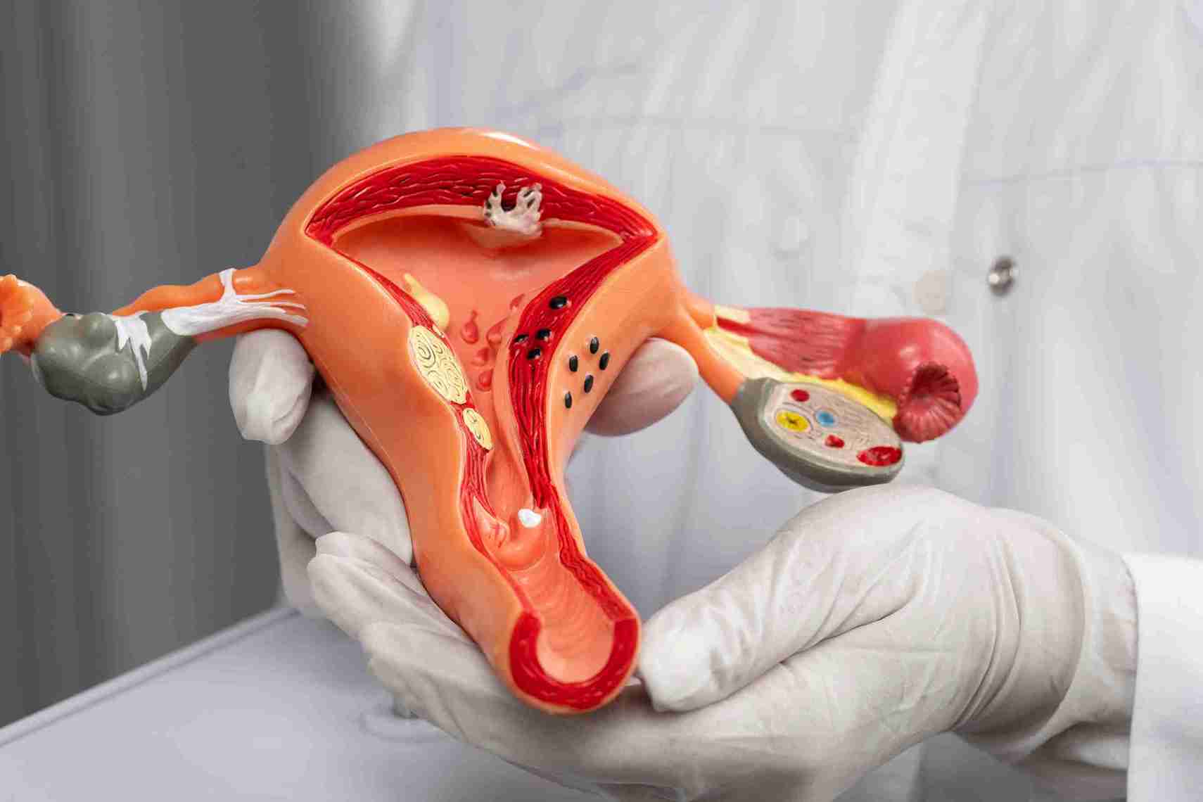
A septated ovarian cyst is a fluid-filled sac in the ovary divided into two or more compartments by internal walls called septa. Multilocular, multiloculated, and septated all describe the same finding. Most are not cancer, particularly before menopause. What matters is not the septa themselves but their thickness, blood flow, and the cyst size.
What is a septated (multilocular) ovarian cyst?
A simple ovarian cyst is one smooth-walled bag of clear fluid. A septated cyst has partitions running across the inside, so instead of one cavity, there are several. Each compartment is called a locule (a single chamber inside a cyst), which is why the same finding is described as multilocular or multiloculated. Radiologists may also write internal septa, internal septations, or loculated cystic lesion. These are wording preferences, not different diagnoses.
The septa form for ordinary reasons in most cases. A benign ovarian tumour called a cystadenoma grows with divisions built into it. Old blood inside a cyst leaves fibrin strands (thin protein threads) that can look like walls. None of this is inherently sinister. Septation describes shape, not behaviour.
Internal walls
None
One or more septa
May or may not have septa
Contents
Clear fluid only (anechoic)
Fluid in two or more compartments
Fluid plus blood, debris or solid tissue
Solid areas
Absent
Usually absent
Often present
Typical risk level
Very low
Low to intermediate, depends on features
Depends entirely on features
Usual next step
Often none needed
Repeat scan, sometimes further tests
Structured risk scoring and specialist review
A septated cyst falls under the broader category of complex cysts, but the reverse isn't true. Not every complex cyst is septated, and a cyst can be complex because it contains blood or solid tissue with no partitions.
Why this one word on a report causes so much worry
Most septated ovarian cysts are found by accident. A woman has a pelvic scan for period pain, a fertility workup, or a routine antenatal check, and the report comes back with a phrase she has never seen. Because the internet in India returns cancer content for almost any query containing "septated" or "multilocular," the leap from a descriptive word to a frightening conclusion happens in about thirty seconds.
The honest position is more measured. Ovarian cysts of some kind affect a large share of women of reproductive age, and the overwhelming majority of septated ones turn out to be benign. Radiologists do not use septation alone to judge risk. They combine it with wall thickness, the presence of solid nodules, blood flow, and your menopausal status, then apply a published scoring system. What happens next depends on that combination, not the single word. If your report flags size rather than architecture, our guide to normal ovary size to get pregnant covers that separately.
Is a septated ovarian cyst cancer? The honest answer
No, in most cases. A septated cyst is far more likely to be a benign cystadenoma, an endometrioma or a resolving haemorrhagic cyst than a cancer, especially before menopause. But septation does move a cyst out of the reassure-and-forget category, so it earns a proper look rather than dismissal.
What the risk figures actually show
Anyone quoting a single percentage for septated cysts is overstating what the evidence supports, because the number changes completely depending on which other features are present. Under the American College of Radiology O-RADS system, a multilocular cyst under 10 cm with smooth inner walls, no solid component, and low blood flow falls into the low-risk band, carrying roughly a 1% to under 10% chance of malignancy. Once the same cyst reaches 10 cm or more, or develops an irregular inner wall or strong blood flow, it moves into the intermediate band, at roughly 10% to under 50%.
Menopausal status shifts the picture again. Before menopause, functional and benign causes dominate, and a multilocular cyst without solid areas is very often a mucinous cystadenoma. After menopause, physiological cysts should not form, so the same appearance is treated with more caution and usually warrants specialist referral regardless of size. This is why a figure copied from an article written for postmenopausal women can badly mislead a woman of thirty.
Thin septations versus thick septations
Thickness is the single most useful detail in your report after size. A septum measuring under about 3 mm is considered thin and is reassuring. A septum measured at 3 mm or more, or described as irregular, nodular or beaded, changes the follow-up pathway. It is important to be clear about what that means: thick does not equal cancer. It means the cyst can no longer be signed off on the scan alone and needs either a repeat study, an MRI, or review by a clinician who specialises in adnexal imaging.
How O-RADS scores a multilocular cyst
O-RADS 1
Normal premenopausal ovary, including follicles and corpus luteum
Effectively nil
No action
O-RADS 2
Almost certainly benign: classic haemorrhagic cyst, typical endometrioma, dermoid, simple cyst under 10 cm
Under 1 percent
Often no follow-up, or one interval scan
O-RADS 3
Multilocular cyst under 10 cm, smooth inner walls, no solid component, colour score under 4
1 percent to under 10 percent
Interval imaging, often by a specialist sonologist
O-RADS 4
Multilocular cyst 10 cm or more, or irregular inner wall or septal irregularity, or any solid component, or colour score 4
10 percent to under 50 percent
MRI or referral to a gynaecologic oncologist
O-RADS 5
Solid mass with strong flow, four or more papillary projections, or ascites and peritoneal deposits
50 percent or more
Gynaecologic oncology referral
Colour score refers to how much blood flow is seen inside the walls and septa on Doppler, graded from 1 (none detectable) to 4 (very strong). It is one of the few features that can shift a cyst between categories on its own.
The IOTA simple rules: B rules and M rules
The International Ovarian Tumour Analysis group publishes ten ultrasound descriptors, five suggesting a benign lesion and five suggesting a malignant one. If only B features are present, the mass is classed benign. If only M features are present, it is classed as malignant. If both appear, or neither does, the result is inconclusive and requires a second-stage assessment. Two of these ten refer directly to multilocular cysts, which is why the rules matter so much for this particular report finding.
Unilocular cyst
Irregular solid tumour
Largest solid component under 7 mm
Ascites (free fluid in the abdomen)
Acoustic shadowing present
Four or more papillary projections
Smooth multilocular tumour under 10 cm
Irregular multilocular solid tumour 10 cm or larger
No detectable blood flow (colour score 1)
Very strong blood flow (colour score 4)
Note the fourth row on each side. A smooth multilocular cyst under 10 cm is benign. The same cyst becomes malignant only when it is irregular, 10 cm or larger, and has solid tissue. The Royal College of Obstetricians and Gynaecologists has incorporated these rules into its guidance for premenopausal ovarian masses.
Why blood flow inside a septum matters more than the septum
True septa carry a blood supply because they are living tissue. Fibrin strands left behind by a bleed do not. This is the practical reason a sonographer runs Doppler across the inside of the cyst rather than only measuring it. Thin, discontinuous strands that do not reach fully from wall to wall and show no flow point strongly towards a resolving haemorrhagic cyst rather than a true multilocular lesion. Thicker linear echoes that cross the cyst completely and light up on Doppler are genuine septations and are assessed differently.
What causes septations to form in an ovarian cyst?
Septa arise through several separate mechanisms, and the mechanism usually tells you more about prognosis than the appearance does.
● Mucinous cystadenoma. The classic multilocular finding. A benign epithelial tumour that grows with many compartments, often filled with thick mucin, and can become very large while remaining entirely benign.
● Serous cystadenoma. Also benign and common, though more often single-chambered than mucinous types. When it does divide, the septa are typically thin and smooth.
● Haemorrhagic cyst. Bleeding into a functional cyst leaves a reticular or fishnet pattern of fine fibrin strands. Patients routinely misread these as septations. They are avascular, do not span the full cyst, and usually resolve within about six to eight weeks.
● Endometrioma. A chocolate cyst arising from endometriosis. Typically shows uniform low-level ground-glass echoes and is often bilocular rather than truly multilocular. Unlike some cysts that resolve on their own, endometriomas usually remain present, making the broader issues of endometriosis and fertility more significant than the cyst itself.
● Dermoid cyst (mature cystic teratoma). Usually single-chambered, but can appear divided because of the mixed tissue types inside, including hair, fat, and calcification.
● Ovarian stimulation for fertility treatment. Gonadotropin stimulation can produce enlarged, multi-compartment-looking ovaries during a cycle. This is expected physiology, not pathology, and is one reason follicular monitoring scans are timed carefully.
● Borderline ovarian tumour. Neither clearly benign nor invasive cancer. May show multilocular architecture with papillary projections and generally requires surgical management.
● Ovarian cancer. The least likely cause in a premenopausal woman, but the one every other finding is measured against. Suggested by thick irregular septa, solid nodules, strong Doppler flow, rapid growth, and free fluid, in combination rather than singly.
Polycystic ovary syndrome does not cause septated cysts. This is one of the most common misconceptions in Indian searches on this topic. The many small follicles seen in PCOS, sometimes described as a string of pearls, are individual antral follicles sitting around the edge of the ovary. They are not one cyst with internal walls, and a PCOS diagnosis neither explains nor excuses a genuine multilocular finding on your report.
What to do after a septated cyst is found
If your report describes benign features
Where the cyst is under 10 cm, the septa are thin and smooth, there is no solid component, and Doppler is quiet, the standard path is watchful waiting with an interval scan rather than immediate intervention. Practical steps:
● Book the repeat scan properly. Ideally just after a period, so a fresh corpus luteum is not mistaken for persistence of the original cyst.
● Ask for the same modality. A transvaginal scan gives far better resolution of internal architecture than an abdominal one, making comparison meaningful.
● Keep the original images, not just the report. Comparison against actual images lets a radiologist say stable rather than probably similar.
● Note any change in symptoms. Increasing girth, persistent bloating or new pain between scans is worth reporting early rather than waiting for the appointment.
If your report describes suspicious features
Thick or irregular septa, solid nodules, papillary projections, strong blood flow, free fluid, or a cyst of 10 cm or more all move you off the watchful waiting path. What follows is escalation of assessment, not automatically surgery:
● Expect further imaging. A pelvic MRI is the usual next step when ultrasound findings are indeterminate, particularly to distinguish an endometrioma from a haemorrhagic cyst or characterise solid areas.
● Expect blood tests in context. Add CA-125 and, in younger women, germ cell markers. These support a risk picture and never make a diagnosis alone.
● Ask which risk score was applied. O-RADS, IOTA simple rules or the Risk of Malignancy Index. Knowing the category is far more useful than knowing the cyst size.
● Ask about referral. If features are genuinely suspicious, the correct referral is to a gynaecologic oncologist, even though that service is outside fertility care. A clinic that routes you appropriately is doing its job.
What does not shrink a septated ovarian cyst?
This section exists because the alternative is you finding the answer somewhere with something to sell.
● Combined oral contraceptive pills do not shrink an existing cyst. A Cochrane review of eight randomised trials covering 686 women found that combined oral contraceptives did not hasten resolution of functional ovarian cysts in any trial. The pill does reduce the formation of new functional cysts, which is a genuine but different benefit.
● No diet dissolves a cyst. Flaxseed, castor oil packs, apple cider vinegar, and anti-inflammatory eating plans have no evidence of shrinking ovarian cysts of any architecture. A septated cyst is a structural finding, not a metabolic one.
● Ayurvedic and homoeopathic preparations have no evidence base here. The preparation itself is rarely the harm. The real issue is the months that pass without an interval scan that would have shown whether the cyst was stable, growing, or resolving. Time is something a septated cyst can genuinely cost you.
● Supplements do not change cyst architecture. No vitamin, herbal extract, or fertility supplement has been shown to alter septations, wall thickness, or vascularity.
● What does help: Attending the interval scan, treating pain adequately, and getting a properly documented risk category from someone who scans adnexal masses regularly will help.
How much does a septated ovarian cyst cost to investigate and treat in India?
Costs split into two stages. The first is evaluation, which almost everyone needs, and which is modest. The second is surgery, which only a minority need. The figures below are indicative ranges rather than quotations, and vary by city, by whether a case is managed as day care or admission, by anaesthesia requirements and by how much workup is done before theatre.
Pelvic or transvaginal ultrasound
₹1,000 to ₹3,500
First line for every septated cyst, and for each interval scan
Pelvic MRI (where indicated)
₹12,000 to ₹19,000
When ultrasound findings are indeterminate or before surgical planning
AMH blood test
₹5,000 to ₹6,500
Ovarian reserve assessment, particularly before any planned cystectomy
Tumour marker panel (where indicated)
₹2,000 to ₹5,000
CA-125 and related markers, guided by age and scan features
Pre-anaesthetic assessment and routine pre-operative bloods
₹4,500 to ₹8,000
Only if surgery is planned
Histopathology of the removed cyst
₹1,500 to ₹4,000
After any surgical removal, to confirm the tissue diagnosis
Laparoscopic ovarian cystectomy
₹80,000 to ₹200,000
Persistent, symptomatic, large or suspicious cysts
Laparotomy (open surgery)
₹35,000 to ₹100,000
Very large cysts, or where the mass must be removed intact
Follicular monitoring scan series
₹1,500 to ₹4,000 per cycle
Where fertility treatment runs alongside cyst surveillance
Diagnostic and pre-operative figures shown here are indicative client-supplied ranges, not audited pricing. Surgical line items remain placeholders pending confirmation. Insurance in India commonly covers ovarian cyst surgery where it is medically indicated, but waiting periods and pre-authorisation requirements apply, and purely elective or diagnostic imaging is often excluded.
When should you see a doctor?
Two different urgencies apply here, and confusing them causes both unnecessary panic and dangerous delay.
Sudden severe one-sided pelvic pain, vomiting, fainting, or fever
Same day emergency care
Possible ovarian torsion or cyst rupture, both time critical
Persistent bloating or abdominal swelling for more than two to three weeks
Appointment within days
Persistent distension is one of the few genuinely useful ovarian red flags
Report describes thick septations, solid areas or strong Doppler flow
Appointment within one to two weeks
Needs structured risk scoring and possibly MRI
Cyst found after menopause, any size or type
Appointment within one to two weeks
Physiological cysts should not form after menopause
Thin septations, under 10 cm, no symptoms
Routine appointment, keep the interval scan
Watchful waiting is appropriate and is not the same as ignoring it
Trying to conceive with a known cyst
Fertility review before starting treatment
Cyst type and size affect timing of stimulation and retrieval
If you are trying to conceive and a scan has flagged a septated or multilocular cyst, a Cloudnine Fertility specialist can review the images alongside your ovarian reserve and plan the sequence properly, rather than treating the cyst and the fertility question as two separate problems. If severe period pain is part of your picture, it is worth reading how painful periods and infertility are connected.
Outcomes: resolution, fertility and pregnancy
Will it go away on its own?
This depends almost entirely on what is causing the septation, which is why a single answer to this question is always wrong.
Haemorrhagic cyst (fibrin strands)
Yes, usually
Six to eight weeks, often within one to two cycles
Functional cyst with strands
Yes, usually
Two to three menstrual cycles
Mucinous or serous cystadenoma
No
Persists and may grow slowly over years
Endometrioma
No
Persists, may enlarge with each cycle
Dermoid cyst
No
Persists indefinitely
Borderline tumour or malignancy
No
Requires surgical management
Does a septated ovarian cyst affect fertility?
For most benign septated cysts, no. Ovulation continues from both ovaries, and the presence of a cyst on one side does not stop the other from working. Women conceive naturally with multilocular cysts in place all the time.
Fertility is affected in specific situations, not as a rule. An endometrioma signals endometriosis, and the underlying disease affects fertility more than the cyst itself. A very large cyst can displace or compress healthy ovarian tissue. Repeated ovarian surgery reduces reserve cumulatively. An awkwardly positioned cyst can also complicate access during egg retrieval.
Will surgery cost me my ovary or my egg reserve?
The ovary is preserved in most benign cases. Laparoscopic cystectomy is specifically designed to shell out the cyst while leaving functioning ovarian tissue behind, and removal of the whole ovary is reserved for situations where preservation is not technically possible, or malignancy is suspected.
The honest caveat, which many clinic pages omit, is that cystectomy does have a measurable cost to ovarian reserve. Systematic reviews and meta-analyses consistently show a fall in AMH after laparoscopic cystectomy, most pronounced for endometriomas, for bilateral surgery, for larger cysts, and where bipolar diathermy is used for haemostasis rather than suturing or haemostatic agents. Some recovery occurs over the following months, though it is often incomplete. This is a reason to test AMH before surgery, not a reason to avoid necessary surgery. Where reserve is already low, and surgery is unavoidable, fertility preservation and egg freezing before the operation is a conversation worth having.
Can you have IVF with a septated ovarian cyst?
Frequently yes, with the sequence planned around the cyst rather than ignoring it. A small benign appearing cyst with thin septa often does not require deferral, and stimulation proceeds with monitoring. A large cyst, one that obstructs the route to the follicles, or one with any suspicious feature is dealt with before a cycle starts, because starting stimulation around an uncharacterised mass is the one genuinely poor option. Where an endometrioma is involved, the decision balances surgical damage to reserve against access and cycle quality, and it goes differently for different people. Which IVF stimulation protocols suit you is decided alongside this, not afterward.
If it is found during pregnancy
Cysts discovered on early pregnancy scans are often corpus luteum cysts that resolve by the second trimester. Persistent multilocular cysts are usually monitored rather than operated on, with intervention reserved for torsion, rupture, obstruction of delivery or genuinely suspicious features. Where surgery is needed electively, the second trimester is generally the preferred window.
Want to consult the Best Fertility in india? Please find the links below.
Find clear, trusted answers to the most common questions about IVF—designed to guide and support you every step of the way.
What does it mean if an ovarian cyst is multiloculated?
It means the cyst has more than one compartment inside it, separated by internal walls. Multiloculated, multilocular, and septated are interchangeable terms for the same finding. It tells you about internal architecture and nothing more. The clinical meaning comes from the additional descriptors alongside it, principally septal thickness, the presence or absence of solid tissue, blood flow on Doppler, and the overall size.
What percentage of septated ovarian cysts are cancerous?
It means the cyst has more than one compartment inside it, separated by internal walls. Multiloculated, multilocular, and septated are interchangeable terms for the same finding. It tells you about internal architecture and nothing more. The clinical meaning comes from the additional descriptors alongside it, principally septal thickness, the presence or absence of solid tissue, blood flow on Doppler, and the overall size.
What percentage of septated ovarian cysts are cancerous?
There is no single honest percentage, because the figure swings with the other features. Using the O-RADS framework, a multilocular cyst under 10 cm with smooth walls, no solid component, and low blood flow sits in a band of roughly 1 percent to under 10 percent. Add a solid component, an irregular inner wall, strong vascularity, or a size of 10 cm or more, and it moves to roughly 10% to under 50%. Menopausal status shifts it further, since benign functional causes are far less likely after menopause.
What is the 1-2-3 rule for ovarian cysts?
It is a naming shorthand used on ultrasound for small, simple, clear structures in the ovary: under 1 cm is called a follicle, roughly 1 cm to 3 cm is a dominant follicle, and above 3 cm it is called a cyst. This rule applies to simple anechoic structures only. It says nothing about a septated cyst, which is categorised by architecture rather than by size alone.