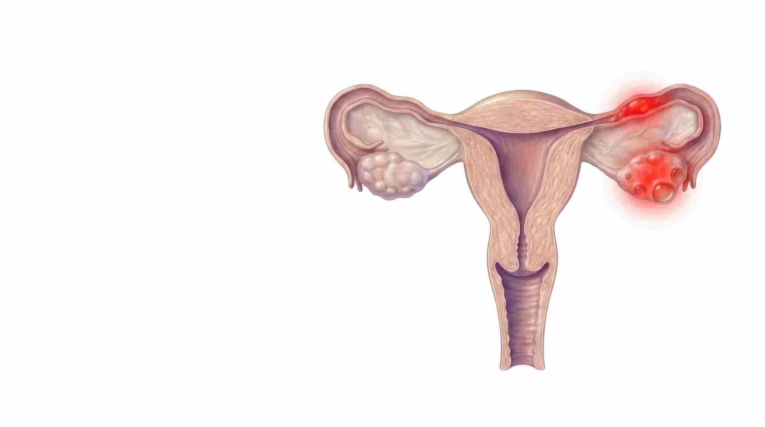
Ovarian cysts are fluid-filled or semi-solid sacs that form on or inside an ovary. The main types are functional cysts (follicular and corpus luteum), haemorrhagic cysts, dermoid cysts, endometriomas, cystadenomas and paraovarian cysts. Most are functional, harmless and resolve on their own within two to three menstrual cycles without any treatment.
What is an ovarian cyst?
An ovarian cyst is a sac of fluid, blood or mixed tissue that develops on or within an ovary. Every woman who ovulates develops a small fluid-filled sac each month, so the presence of a sac on a scan is usually normal physiology rather than disease. Cysts are grouped into two broad families. Functional cysts arise from the ordinary hormonal work of the menstrual cycle and disappear by themselves. Pathological cysts arise from abnormal cell growth or from another condition such as endometriosis, and they behave differently.
Feature
Functional Cysts
Pathological Cysts
Origin
Related to normal ovulation and menstrual-cycle hormones.
Related to abnormal cell growth or an underlying ovarian condition.
Typical Age
Most common during the reproductive years, before menopause.
Can occur at any age, including after menopause.
Usual Course
Usually resolves spontaneously within two to three cycles.
May persist and can gradually enlarge depending on the type.
Usual Treatment
Usually observation and follow-up when clinically appropriate.
Depends on the type, size, ultrasound appearance, symptoms, and other clinical factors.
Examples
Follicular cyst and corpus luteum cyst.
Dermoid cyst, endometrioma, and cystadenoma.
Why ovarian cysts are found so often in India
Most ovarian cysts in India are discovered by accident. Pelvic ultrasound has become inexpensive and widely available, and it is now routinely performed during master health checkups, antenatal visits, abdominal pain assessments and fertility workups. A scan that would once never have been done now finds a two-centimetre sac that was always there and would always have vanished on its own.
This matters because the finding often causes more distress than the cyst does. A woman handed a report that reads "left ovarian cyst" frequently assumes surgery or cancer. In reproductive-age women, the overwhelming majority of these findings are physiological, and international radiology consensus treats follicles up to three centimetres in premenopausal women as an entirely normal ovary rather than a cyst. The useful question is never simply whether a cyst is present. It is which type it is, how it looks on the scan, and whether it is causing any problem.
What are the types of ovarian cysts?
Ovarian cysts fall into two groups: functional cysts, which are a by-product of ovulation, and pathological or other cysts, which have a separate cause. The sections below cover every type you are likely to see named on an Indian ultrasound report.
Functional cysts
Functional cysts are by far the most common type and form as part of the normal menstrual cycle. They are benign, usually painless, and typically disappear within two to three cycles without treatment.
Follicular cyst. Each month, a follicle (the fluid-filled sac holding an egg) grows and then ruptures to release the egg. If it fails to rupture, it keeps filling with fluid and becomes a follicular cyst. These are usually silent, found incidentally, and resolve without intervention.
Corpus luteum cyst. After the egg is released, the empty follicle becomes the corpus luteum, a temporary hormone-producing structure. If it seals over and fills with fluid or blood instead of shrinking, a corpus luteum cyst forms. These may cause one-sided pelvic ache, a delayed period or light spotting, and they can be a normal finding in early pregnancy, where they support the pregnancy until the placenta takes over.
Haemorrhagic cyst
A haemorrhagic cyst is a functional cyst in which a small blood vessel in the cyst wall bleeds, filling the sac with blood. It classically causes sudden, sharp, one-sided lower abdominal pain, sometimes triggered by exercise or intercourse. On ultrasound, it has a distinctive lace-like or cobweb internal pattern that a trained sonographer can recognise. Despite the alarming name and the pain, most haemorrhagic cysts settle without surgery, and a repeat scan after six to eight weeks, timed to a different phase of the cycle, usually shows resolution.
Dermoid cyst (mature cystic teratoma)
Dermoid cysts arise from germ cells, the cells that would ordinarily become eggs, and because those cells can form any body tissue, a dermoid may contain hair, skin, fat, teeth or even bone. They are benign, grow slowly, and are among the most common ovarian tumours found in young women. Their significance is mechanical rather than cancerous: because they are dense and heavy, larger dermoids carry a raised risk of ovarian torsion, in which the ovary twists on its blood supply. That is a surgical emergency. Removal is usually advised for dermoids that are large, growing, or causing symptoms, and the aim is always to shell out the cyst while preserving the healthy ovary around it.
Endometrioma (chocolate cyst)
An endometrioma forms when endometriosis, a condition in which tissue similar to the uterine lining grows outside the uterus, involves the ovary. Each cycle that tissue bleeds, and because the blood has nowhere to drain, it collects and darkens, giving the thick brown contents that earn the name chocolate cyst. Endometriomas typically cause severe period pain, chronic pelvic pain, pain during intercourse and difficulty conceiving.
Of all the cyst types, this is the one with the most direct fertility consequences, and also the one where treatment decisions are most often got wrong. It is covered in detail further down.
Cystadenoma
Cystadenomas are benign tumours that grow from the cells on the outer surface of the ovary. Unlike functional cysts, they do not resolve on their own, and a simple-looking cyst that steadily grows over successive scans is most likely to be a cystadenoma. There are two forms. Serous cystadenomas are filled with thin watery fluid and usually stay moderate in size. Mucinous cystadenomas are filled with thick mucus-like fluid and can become genuinely large, occasionally filling much of the abdomen before they are found. Both are usually removed surgically once they reach a size that causes pressure symptoms or diagnostic uncertainty.
Polycystic ovaries in PCOS (not true cysts)
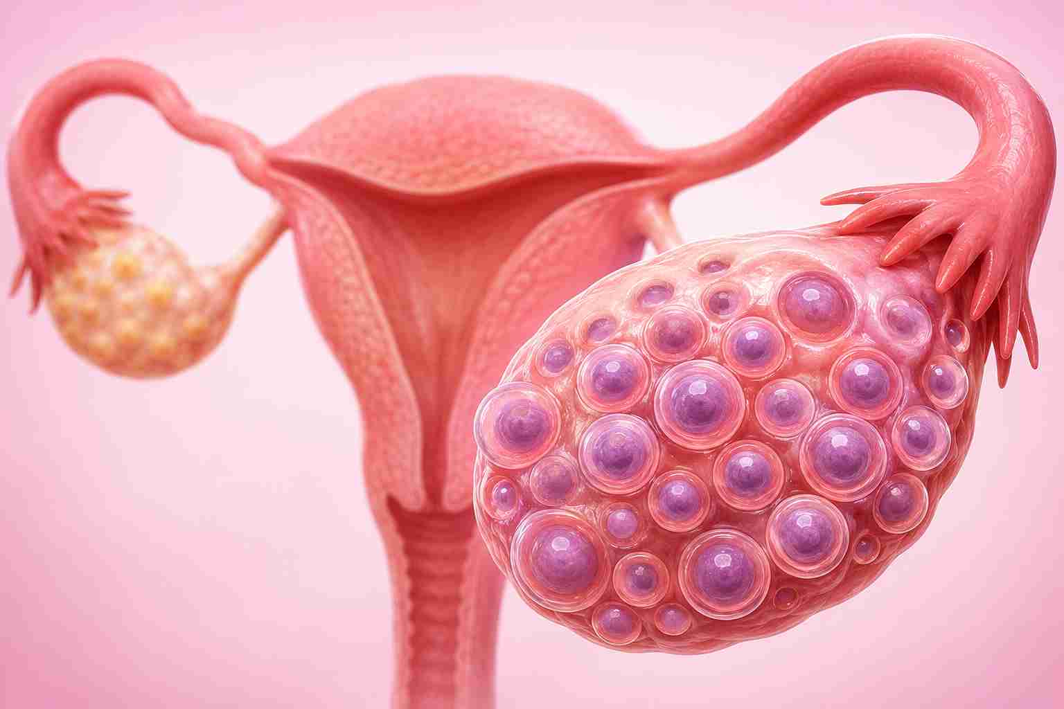
This is the single most misunderstood entry on any ovarian cyst list. The multiple small structures seen around the ovary in Polycystic Ovary Syndrome are not cysts. They are immature antral follicles that have stalled because ovulation is not happening regularly. They do not rupture, they do not bleed, they do not twist, and they are never removed surgically.
The 2023 International Evidence-based Guideline defines polycystic ovarian morphology as twenty or more follicles of two to nine millimetres in at least one ovary, or an ovarian volume of ten millilitres or more. That guideline also now allows a raised AMH (anti-Mullerian hormone) blood level to be used instead of ultrasound in adults, and it specifically advises against using either method to diagnose the condition in adolescents. Crucially, polycystic ovarian morphology alone is not PCOS. The diagnosis needs two of three features: irregular ovulation, clinical or biochemical signs of raised androgens, and polycystic morphology. Many women with entirely normal cycles have polycystic-looking ovaries and no syndrome at all.
Paraovarian and inclusion cysts
A paraovarian cyst develops in the tissue beside the ovary rather than in it, arising from embryonic remnants in the broad ligament. It is benign, does not respond to cycle hormones, and therefore does not resolve, but small ones need nothing more than observation. An ovarian inclusion cyst is a tiny sac formed when surface tissue is trapped within the ovary after ovulation, infection or previous pelvic surgery. These are almost always incidental and asymptomatic.
Complex ovarian cyst
Complex is a description on a scan report rather than a diagnosis. It means the sac is not uniformly filled with clear fluid: it contains solid areas, internal walls called septations, or mixed contents. A haemorrhagic cyst, a dermoid and an endometrioma will all read as complex, and all three are benign. So the label alone is not a reason for alarm. It is, however, a reason for proper characterisation, because a small minority of complex cysts represent a borderline or malignant tumour. Depending on the appearance and your age, the next step may be a repeat scan at a different point in the cycle, an MRI, or a CA-125 blood test in selected patients. CA-125 is used with care in premenopausal women because endometriosis, fibroids, infection and even menstruation itself can raise it without any cancer being present.
Which types of ovarian cyst affect fertility?
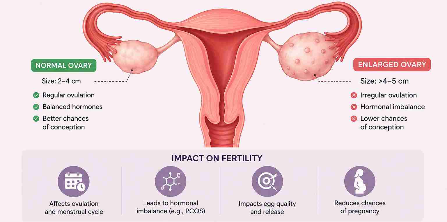
Most do not. Functional, haemorrhagic, paraovarian and inclusion cysts have no meaningful effect on your chance of conceiving, and neither do most small dermoids or cystadenomas. Three situations genuinely matter. Endometriomas reduce fertility through the endometriosis that causes them, through inflammation, and through damage to the surrounding ovarian tissue. Polycystic ovaries in PCOS affect fertility because ovulation is irregular, not because of the follicles themselves. Any cyst large enough to distort pelvic anatomy or obstruct access to the ovary during egg collection can complicate treatment. Everything else is usually a bystander.
Different cyst types have entirely different causes, which is why no single preventive measure works for all of them.
Normal ovulation. The commonest cause by far. A follicle that does not rupture, or one that seals over after releasing the egg, becomes a functional cyst. This is biology working slightly out of step, not a disease.
Hormonal imbalance. When ovulation is irregular, follicles accumulate without maturing. This underlies the polycystic ovarian appearance seen in PCOS.
Endometriosis. Tissue similar to the uterine lining implants on the ovary, bleeds each cycle, and forms an endometrioma.
Fertility treatment. Ovulation induction and IVF stimulation deliberately grow multiple follicles, so functional cysts are a common and expected finding during and just after a treatment cycle.
Germ cell development. Dermoid cysts arise when a germ cell begins forming mixed body tissues. This is not caused by anything you did or ate.
Pregnancy. A corpus luteum cyst supporting an early pregnancy is normal and usually resolves by the second trimester.
Pelvic infection or previous surgery. Inflammation and scarring can trap tissue and produce inclusion cysts or adhesion-related collections.
There is no credible evidence that diet, weight, cold foods, sitting posture or stress cause ovarian cysts, despite how commonly these are blamed.
What to do if a scan shows an ovarian cyst
The right next step depends almost entirely on how the cyst looks and how big it is, not on whether one is present.
If the cyst is simple and small
A simple cyst is thin-walled, round, and filled with clear fluid, with no solid areas or septations. In premenopausal women, these are managed by watchful waiting.
Up to 3 cm. Considered a normal ovary. Radiology consensus does not require this to be reported as a cyst, and no follow-up is needed.
3 cm to 5 cm. Almost certainly physiological. No routine follow-up imaging is required. A repeat scan after two to three cycles is reasonable if you have symptoms.
5 cm to 7 cm. Annual ultrasound follow-up is generally recommended so that any growth is picked up.
Above 7 cm. Further imaging such as MRI, or a surgical opinion, because the whole cyst can be difficult to characterise fully on ultrasound alone.
One important point on timing: a repeat scan is far more informative if it is done in the first week after your period, when a physiological cyst will have collapsed. A scan repeated in the same phase of the cycle can show a fresh follicle and be misread as a persistent cyst.
If the cyst is complex, large or persistent
Here the goal is characterisation before any decision about surgery. Expect a detailed transvaginal ultrasound performed by an operator experienced in adnexal masses, which is the single highest yield test available. MRI is added when the ultrasound appearance is uncertain. CA-125 is used selectively, weighted by your age and menopausal status, because it is much less reliable before menopause. If surgery is needed, laparoscopy (keyhole surgery) is the standard approach for benign cysts, and cystectomy, meaning removal of the cyst while conserving the ovary, is strongly preferred over removing the whole ovary in women who may want to conceive. Ask directly whether ovarian preservation is planned, because it is not always the default.
A word on what does not work. A Cochrane review of eight randomised trials covering 686 women found that combined oral contraceptives do not speed up the resolution of functional ovarian cysts, whether the cysts arose spontaneously or after ovulation induction. Pills are useful for preventing new functional cysts from forming, and for managing pain, but they will not shrink the cyst you already have. Watchful waiting for two to three cycles is the appropriate approach, and a cyst that persists beyond that is more likely to be pathological and to warrant a surgical opinion.
How much does ovarian cyst diagnosis and surgery cost in India?
Costs vary by city, by whether the procedure is done as day care or with admission, by anaesthesia, and by how much work is needed if endometriosis or adhesions are found during surgery. The figures below are drawn from the Cloudnine Fertility audited pricing sheet where the item is covered.
Test or Procedure
Indicative Cost Range
Pelvic or Transvaginal Ultrasound
₹1,000–₹3,500
Follicular Monitoring Scans
₹2,800–₹3,500
AMH Blood Test
₹5,000–₹6,500
CA-125 Blood Test
₹2,000–₹5,000
Pelvic MRI
₹12,000–₹19,000
Diagnostic Laparoscopy
₹30,000–₹75,000
Laparoscopic Ovarian Cystectomy
₹75,000–₹1,50,000
Egg Freezing Before Ovarian Surgery
~₹82,500, including three months of storage, plus consumables
IVF or ICSI Cycle
₹1.15 lakh–₹2.5 lakh all-in, plus stimulation injections of ₹70,000–₹95,000
When to see a doctor about an ovarian cyst
Most cysts need patience rather than treatment. A small number need attention the same day, because a cyst can rupture or the ovary can twist on its blood supply, and both are time-critical.
What You Notice
What to Do
Sudden severe one-sided pelvic pain, with vomiting, faintness
Go to an emergency department the same day. This can indicate ovarian torsion or cyst rupture.
Pelvic pain that is persistent but not severe
Book a gynaecology appointment within a week or two for assessment and, if appropriate, an ultrasound.
Period pain severe enough to interfere with work or study, with pain during intercourse
Seek assessment for possible endometriosis rather than assuming it is normal period pain.
A cyst reported as complex, or one that has grown between two scans
Ask for specialist characterisation before agreeing to any surgery.
Irregular or absent periods with a polycystic ovarian appearance
Ask for a PCOS assessment. Polycystic-appearing ovaries alone do not necessarily require treatment.
Trying to conceive for a year and a cyst has been found
Book a fertility evaluation so the cyst can be assessed alongside the overall fertility picture.
If a scan has found an ovarian cyst and you are trying to conceive, or planning to in the next few years, you can book a consultation with a Cloudnine Fertility specialist to have the cyst assessed in the context of your ovarian reserve and your timeline, rather than in isolation.
What outcomes can you expect by cyst type?
The realistic outlook differs sharply across types, and knowing which one you have tells you most of what you need to know.
Cyst Type
Resolves Alone
Surgery Usually Needed
Effect on Fertility
Follicular
Yes, often within two to three cycles
No
Usually none
Corpus Luteum
Yes, often within two to three cycles
No
Usually none
Haemorrhagic
Usually within six to eight weeks
Rarely, mainly if it ruptures with significant bleeding
Usually none
Dermoid
No
May be considered if large, growing, or symptomatic
Usually minimal if ovarian tissue is conserved
Endometrioma
No
Case by case; surgery is not routine for every endometrioma
Can affect fertility and may reduce ovarian reserve, particularly with some surgical approaches
Cystadenoma
No
Usually considered when symptomatic or large
Usually minimal if the ovary is conserved
Polycystic Ovaries
Not applicable
Not usually treated with surgery
May affect fertility through irregular or absent ovulation
Paraovarian
No
Usually only if large, symptomatic, or causing complications
There is no 3:2:1 rule in any major gynaecology or radiology guidelines. The term appears to be a recently circulated online aide-mémoire rather than an established clinical standard. A separate 1-2-3 rule is sometimes used in ultrasound terminology to describe follicle and cyst size. However, clinical management depends on the size, appearance and other features of the cyst, based on established radiology and gynaecology guidelines.
What are the four types of ovarian cysts?
When sources refer to four types, they usually mean functional cysts, dermoid cysts, endometriomas and cystadenomas. That shortlist is a reasonable summary of the most common presentations, but it is incomplete. It leaves out haemorrhagic cysts, which are a frequent cause of sudden pelvic pain, and paraovarian cysts, which sit beside the ovary rather than in it. It also tends to include polycystic ovaries as a fifth type, which is inaccurate, because those are immature follicles rather than cysts.
What size of ovarian cyst is dangerous?
Size alone is a weaker signal than appearance. A ten centimetre simple cyst is usually benign, while a four centimetre cyst with solid nodules and blood flow needs urgent characterisation. That said, size does carry practical weight. In premenopausal women, simple cysts up to five centimetres need no follow-up, five to seven centimetres warrant annual scans, and above seven centimetres further imaging or a surgical opinion is appropriate. Larger cysts of any type also carry a higher risk of torsion. After menopause, the thresholds are lower, and any solid component is taken more seriously.
Can Ayurveda cure an ovarian cyst?
No published clinical trial evidence shows that any Ayurvedic, homoeopathic, or herbal preparation dissolves an ovarian cyst. The reason these remedies appear to work so often is that the great majority of cysts are functional and would have resolved within two to three cycles regardless of what was taken during that window. The real risk is delay. A dermoid, a cystadenoma or an endometrioma will not resolve, and months spent on an alternative remedy are months in which a torsion risk goes unaddressed or ovarian reserve quietly declines. If you wish to use traditional medicine alongside conventional care, tell your gynaecologist, and do not let it replace the follow-up scan.
Schedule your consultation Today!
Book your consultation with our fertility experts the first step toward your parenthood journey.